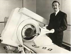
Computed Tomography (CT) imaging is also known as "CAT scanning" (Computed Axial Tomography). CT was invented in 1972 by British engineer Godfrey Hounsfield of EMI Laboratories, England and by South Africa-born physicist Allan Cormack of Tufts University, Massachusetts (Imaginis, 2007). They were both awarded the Nobel Prize in 1979 (EMedicineHealth, 2010). The first CT scanners were used between 1974 and 1976. Originally, the systems were only dedicated to head imaging, but systems that could scan the whole body were available in 1976. CT became known and became widely available by about 1980. There are now about 6,000 CT scanners installed in the U.S. and about 30,000 installed worldwide (Imaginis, 2007).
The first CT scanner developed took a few hours to get the raw data for a single scan or "slice" and took days to reconstruct a single image from this raw data. Since the time CT was developed, it has made great developments in speed, patient comfort, and resolution. Today, more body parts are scanned in less time. Faster scanning helps to eliminate artifacts from patient motion such as breathing or peristalsis. The scans are now faster and more patient-friendly than ever before. Overwhelming research and improvements have been done to provide excellent image quality for diagnostic confidence at the lowest possible x-ray dose (Imaginis, 2007).