Shaileen
Ejtemai—Final Project
Part I - Choosing a
Protein-Hetero Compound Complex
This project is about relation of the protein with its hetero compound; the protein chosen randomly from HIC-Up website. The protein is hydrolase (PDB code1LI6) and its hetreo compound 5- Methylpyrrole (5MP) with formula of C5 H7 N. 1LI6 is a T4 lysozyme mutanted (l99a/m102q). It is found in bacteriophage t4. In biochemistry, a hydrolase is an enzyme that catalyzes the hydrolysis of a chemical bond; causing a splitting of chemical bond with the addition of the elements of water. An interesting observation is that, 5MP can not be the name of the compound, because the structure shows a Methylpyrrole. In other words, 5-MP does not exist. On the other hand, all the protein related databases and researches, mention the compound under 5-MP. 1LI6 function is hydrolysis of the 1, 4-beta-linkages between n- acetyl-d-glucosamine and n-acetylmuramic acid in peptidoglycan heteropolymers of the prokaryotes cell walls.
Part II - Extracting
the Hetero Compound
Table1. Energy
comparison before and after MM2.
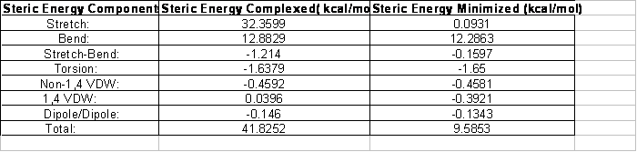
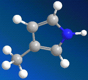
Figure 1. 3D Structure of 5-Methylpuyrrole
after Energy Minimization
Compounds complexed within a protein are rarely in their lowest energy state. Using Chem3D software, the steric energy of the hetero compound was calculated before and after energy minimization. MM2 caused a huge decrease in total steric energy. The initial value indicates the steric energy of the hetero compound complexed with hydrolase. The after MM2 steric energy shows the energy of the hetero compound free of intermolecular interactions and in gas phase. When compound is complexed with protein, each of components of steric energy (mentioned earlier in Table 1), have different and usually higher values. These energy components, cause changes in protein bonds. As it can be seen, energy minimization has caused decrease in all of the energies; More significant reductions in some energy. The process of the geometry optimization is to optimize the total energy with respect to the nuclear coordinates.
Stretch term is
to show whenever a bond is compressed or stretched the energy goes up (term
Stretch-bend term explains the changes in energy as angles are bent from their norm and the energy increases.
When a bond angle is reduced the two bonds forming the angle will stretch to alleviate the strain
Intermolecular rotations (rotations about torsion or dihedral angles) require energy (Torsion strain).There was no significant change in this energy, this could have happened as none of the bond angles were changed significantly. The van der Waals radius of an atom is its effective size. As two non-bonded atoms are brought together the van der Waals attraction between them increases (a decrease in energy); this is called van der Waals Interactions. Here they changes slightly.
Dipole/dipole gives the energy associated with the bond’s dipole interaction. Here the change caused by MM2 is very insignificant.
Part III -
Superimposing the Extracted and the Energy-Minimized Hetero Compounds
As seen in Table 1, there is a large
difference between the resulted total steric energies. However, overlaying the
structures of before and after MM2 does not support this. The reason for this
could be that Fast Overlay from Chem 3D software. In this process the fragment
of before energy minimization was used as the target fragment and the other
structure was imposed on it. The table 1. Shows that the most difference in
total energies is caused by stretch energy. This means that the length of bonds
have decreased after energy minimization; this is caused as protein’s amino
acids do not attract the compounds atoms and bonds. Therefore, the van der
Waals energy decreases too.
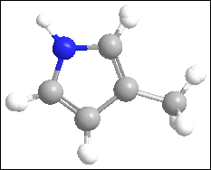
Figure 2.Overlay of
Extracted and Energy Minimized hetero compound.
Part IV -
Protein-Ligand Interactions
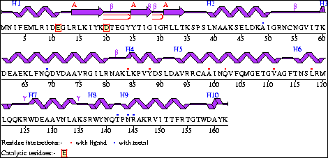
Figure 3: Wire
Diagram of Hydrolase
The wiring diagram was from the
Protein Data Bank website. This diagram helps in understanding the primary and
secondary structure of the 1li6 protein. The red dots in diagram shows
which amino acid residues interact with the hetero compound.
.
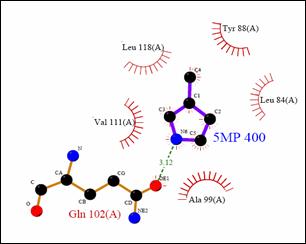
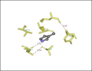
Figure
4: Ligplot Compared to DS Viewer Image of 1LI6
protein Interactions with 5-Methylpyrrole
This table shows the interactions between Amino
acid residues and the 5-Methylpyrrole. As it can be seen the interactions by
DSviewer can be seen in actual 3D, compare to 2D of the ligplot.
Table2. Amino Acid Ligand
Interactions
|
Amino
acid |
Type |
Atoms
of Attraction |
Type
of interaction |
|
Leu 118 |
non polar |
CH |
Hydrophobic |
|
tyr 88 |
non polar
-aromatic |
CH |
Hydrophobic |
|
Leu 84 |
non polar |
CH |
Van der
Waals |
|
Val 111 |
non polar |
CH |
Van der
Waals |
|
Gln 102 |
polar,
uncharged |
N |
Hydrogen bond |
|
Ala 99 |
non polar |
CH |
Van der
Waals |
To find the amino acid residues that interact with hetero compound, figure3. The wire diagram was used. Then these residues were selected in proDSviewer.
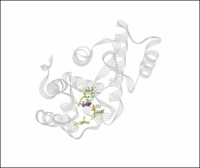
Figure 6: Hetro compound Complexed with protein, showing the interacting amino acid residues
The complete interactions within the protein are shown in Figure 6. Here the interacting amino acids are yellow stick models, the hetero compound is gray and blue ball and stick model, and the rest of the protein is shown in a line ribbon model.
Part V - Bibliographic Information
1. Yamaguchi,
K. Iida, N. Matsui, S. Tomoda, K. Yura, M. Go: Het-PDB Navi. : A database for
protein-small molecule interactions. J. Biochem (
2. Wei, B.Q., Baase, W.A., Weaver, L.H., Matthews, B.W., Shoichet, B.K. A Model Binding Site for Testing Scoring Functions in Molecular Docking J.Mol.Biol. v322 pp.339-355 , 2002 http://www.ncbi.nlm.nih.gov/entrez/query.fcgi?cmd=Retrieve&db=PubMed&dopt=Abstract&list_uids=12217695
3. Kleywegt, G.J. and Jones, T.A. (1998). Databases in protein crystallography. Acta Cryst D54, 1119-1131 (CCP4 Proceedings)http://xray.bmc.uu.se/hicup/
4. H.M. Berman, J. Westbrook, Z. Feng, G. Gilliland, T.N. Bhat, H. Weissig, I.N. Shindyalov, P.E. Bourne: The Protein Data Bank. Nucleic Acids Research, 28 pp. 235-242 (2000) http://www.rcsb.org/pdb/navbarsearch.do?newSearch=yes&isAuthorSearch=no&radioset=All&inputQuickSearch=1li6&image.x=0&image.y=0ℑ=Search