::  A N C H A   L A B  ::
:: WELCOME
:: FOCUS OF THE LAB
:: METHODS
:: PAPERS
:: COURSES
:: JOURNAL CLUB
:: PEOPLE
:: AWARDS
:: CONTACT ME
:: LAB ALUMNI
 

Ectopic synthesis of melanin in human adipose tissue may prevent secondary complications of obesity

Manpreet Randhawa, Sandy Page, Zobair Younossi, Vikas Chandhoke, Vincent J. Hearing, Ancha Baranova

This is a collaborative project between:

School of Systems Biology, College of Science, George Mason University, Fairfax, VA

Translational Reseach Institute, Inova Hospital, VA

Pigment Cell Division, NCI, NIH

Preliminary results for this study were published in FASEB J. 2009 Mar;23(3):835-43.

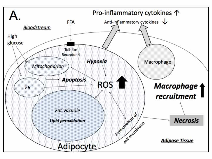
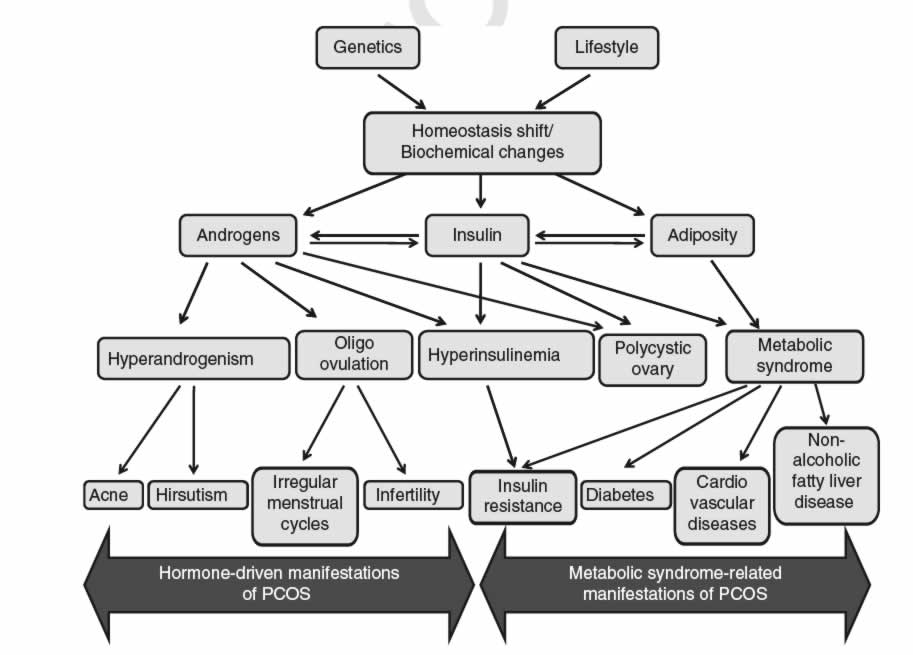
Obesity has been strongly associated with systemic inflammation and, to a lesser degree, with oxidative stress, although the causal relationships among these factors are unclear. Our recent study demonstrated an expression of the components of the melanogenic pathway and the presence of melanin in visceral adipose has raised questions regarding the possible role of melanogenesis in adipose tissue. We also found larger amounts of melanin in the adipose tissue of obese patients relative to lean ones. We hypothesize that melanin, a pigment known for its antioxidant and anti-inflammatory properties, may scavenge reactive oxygen species and abate oxidative stress and inflammation in adipose tissue. It is possible that the α-melanocyte-stimulating hormone or its synthetic analogues could be used to stimulate melanin production in adipocytes and, by that, to prevent the development of the secondary complications of obesity, namely, Non-Alcoholic Fatty Liver Disease, Metabolic Syndrome, Cardiovascular conditions and PCOS.


This research has been supported by a grant from the Thomas F. Jeffress and Kate Miller Jeffress Memorial Trust and by the Intramural Research Program of the National Cancer Institute at NIH.

The details of the hypothesis outlined above could be found in our recent paper published by Ovesity Review "Melanin and melanogenesis in adipose tissue: possible mechanisms for abating oxidative stress and inflammation?"

Press-releases associated with this direction of our research can be downloaded from here:

FASEB Journal press release:

GMU press-release:

This paper has been also reviewed by Prof. Shosuke Ito in the Commentary: Melanin seem to be everywhere in the body, but for what? published in Pigment Cell Melanoma Res. 2009 Feb;22(1):12-3.

This Figure describe the mechanism of how the melanin produced in adipocytes abates oxidative damage and release of pro-inflammatory molecules.

melanin mechanism

mechanism 2

FIGURES from the manuscript published in FASEB Journal

Figure 1 and Figure 2.

Figure 3.

Figure 1. Fontana-Masson stain of human adipose tissue demonstrates melanin pigment (black staining) mainly in the periphery of the adipocytes.
A and B. Multiple conglomerates of melanin granules are present at the periphery of the adipocytes in adipose tissue from morbidly obese subjects (20x magnification).
C and D. Melanin granules are scarce in the adipocytes of adipose tissue from non-obese subjects (20x magnification).
E. No melanin granules were observed in the microvessels located in the adipose tissue (20x magnification).
D. Melanin staining in skin tissue used as a positive control (10x magnification).

Figure 2. Immunohistochemical staining of visceral adipose tissue sections from morbidly obese and from non-obese subjects for human TYR, TYRP1 and TYRP2 proteins (20x magnification). Red: TYR, TYRP1 or TYRP2 staining. Blue: DAPI (nuclei).
A, C, E, G: Visceral adipose tissue from a morbidly obese subject.
B, D, F, H: Visceral adipose tissue from a non-obese subject.
A and B: Tyrosinase;
C and D: Tyrosinase-related protein Tyrp2 (dopachrome tautomerase);
E and F: Tyrosinase-related protein Tyrp1; G and H: Negative control (secondary antibodies) and DAPI.

Figure 3. The presence of melanin in extracts of adipose tissue as revealed by LC-UV-MS.
A. Homogenized samples of adipose tissue separated into three phases: supernatant (fat), aqueous and sediment. Cell debris sediments from adipose tissue samples from morbidly obese subjects contain visible amounts of black pigment. a and b: adipose sediments of non-obese subjects; c and d: adipose sediments from obese subjects.
B: The CAD of the PTCA precursor ion at mass-to-charge ratio (m/z) 198 produces abundant product ions at m/z 154 and 110 peaks.
C. LC-MS Multi-ion SIM chromatogram of PTCA peak at a retention time of 6 min.
D. Negative ESI mass spectra of PTCA peak at 6 min. E. HPLC – UV/VIS Chromatograms at 270 nm.